# The Nasolacrimal System
# Anatomy and Physiology
The tear film components are produced by the lacrimal gland (aqueous), gland of the third eyelid (aqueous), tarsal/Meibomian glands (lipid) and goblet cells (mucin).
When the eyelids blink the tears are pushed from lateral to medial so they can go down the lacrimal puncta. From the puncta the tears drain down the canaliculi, into the lacrimal sac, down the nasolacrimal duct to exit the nose or the pharynx.
# Epiphora
Epiphora is excessive tearing outside of the nasolacrimal system which can be considered a result of either excessive production or from decreased outflow from the nasolacrimal system.
Reflex secretion: Secretion that occurs secondary to mechanical irritation from hairs or foreign bodies, painful ocular diseases (ulcers, uveitis), or irritants such as light and wind.
- Treatment: Determine and treat primary cause
Small punctum
- Treatment
- Enlarge the opening using topical anesthetic and an 11 blade
- Treatment
Imperforate punctum
- Treatment
- Create an opening with scissors. This needs to be done under general anesthesia. Then treat medically with topical antibiotic/steroid ointment TID-QID for one week to try to prevent reclosure.
- Treatment
# Dacryocystitis
Primary infection or infection secondary to presence of foreign body in the duct or lacrimal sac.
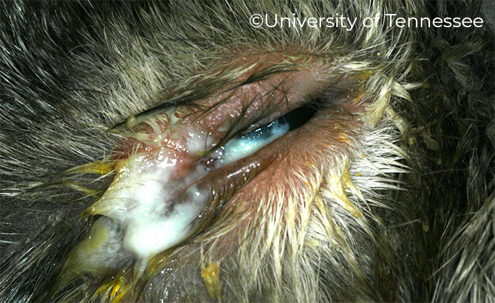
- Relatively rare condition in dogs and cats
- Most common clinical finding is excessive purulent discharge from the medical canthal area (where the lacrimal puncta/ducts are)
- Usually the globe is minimally affected and the conjunctivitis is mild-moderate
Diagnosis
- Thick exudate at the medial canthus
- The amount of exudate is excessive for the degree of conjunctivitis
- Air bubbles may be present
- Pain or swelling may be present at the medial canthus
- Cornea is normal The discharge may be streaked with normal tears
- Dachryocystorhinography may be helpful to localize the blockage
Treatment
- Attempt to flush the duct, do aerobic and anaerobic cultures. With acute disease treat with topical AB/steroid solution and systemic antibiotics for 6 weeks with periodic flushes. With chronic disease an indwelling catheter may be necessary. Surgery may be necessary if the infection is secondary to a foreign body.
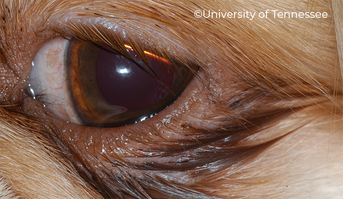
# Tear staining
Staining of the hair at the medial canthus is seen primarily in poodles, spitz and other small white dogs. Multiple conditions contribute to this phenomenon: medial aberrant dermis, abnormal lower punctum, medial lower entropion, prominent eye with shallow lacrimal lake, distichia or trichiasis causing reflex tearing
Treatment
- Very difficult to resolve. Systemic tetracycline or metronidazole may give a temporary fix but long-term therapy is frowned upon. Surgical procedures may or may not be of benefit.
← Eyelids KCS/Dry Eye →