Lens diseases manifest as an abnormal appearance or an abnormal location of the lens. Vitreous diseases manifest primarily as an abnormal appearance.
# Cataract
Definition : An opacity of the lens of the eye. If severe enough, will prevent light from reaching the retina and therefore impair vision.
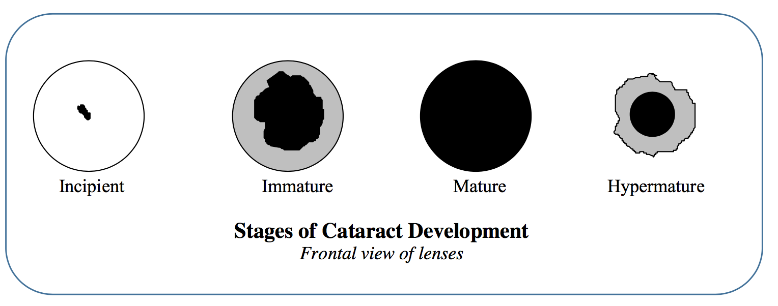
Classification based on “stage of development” (i.e., degree of lens involvement)
Incipient - Small focal opacity
Immature - More diffuse, but fundic reflection still easy to see
Mature - Fundic reflex barely visible or completely obliterated
Hypermature - Lens material liquifies and leaks out of lens capsule into the interior of the eye. Major sign that cataract is hypermature is wrinkling of lens capsule. In addition, the lens material in hypermature cataracts often appears “glittery.” Fundic reflex may or may not be visible depending on how much lens material has leaked out. Usually associated with anterior uveitis of varying degree.
Cataracts usually (but not always!) progress from incipient through mature or hypermature stages.
# Etiologies
Genetic. Documented in numerous breeds
Documented in numerous breeds
Goldens and Huskies
- Characteristic posterior subcapsular opacity
Diabetes mellitus. Glucose levels in lens exceed capacity of glycolytic pathway, the excess is shunted toward production of that can’t escape the lens (sorbitol, fructose). Result is hyperosmotic lens that imbibes water, swells significantly (“intumescence”) and opacifies.
- The first enzyme in that sorbitol pathway is aldose reductase. As such, it would make sense that if AR could be inhibited then progression down the sorbitol pathway would not be possible. An AR inhibiting eyedrop is indeed in development.
Secondary to uveitis. Is seen in all species, and is the most common cause of cataracts in horses and cats.
Nutritional cataract. Cataracts are relatively commonly seen in orphaned animals raised on milk replacer. Arginine is often blamed as the missing nutrient but there is little actual evidence. As the animal ages the “new” lens material is usually normal, so nutritional cataracts may cause minimal symptoms in the adult.
Age related cataract. Cataracts due to age, which are the rule in humans, really aren’t that common in dogs. When they do occur they are often most prominent in the nucleus.
Cataracts associated with Progressive Retinal Atrophy (PRA). PRA is a genetic retinal disease that causes irrevocable (and untreatable) blindness. We’ll study PRA in detail later in the term. A certain percentage of PRA dogs have concurrent cataracts for reasons that aren’t entirely clear. The importance of knowing this is that cataract removal won’t improve vision in these dogs.
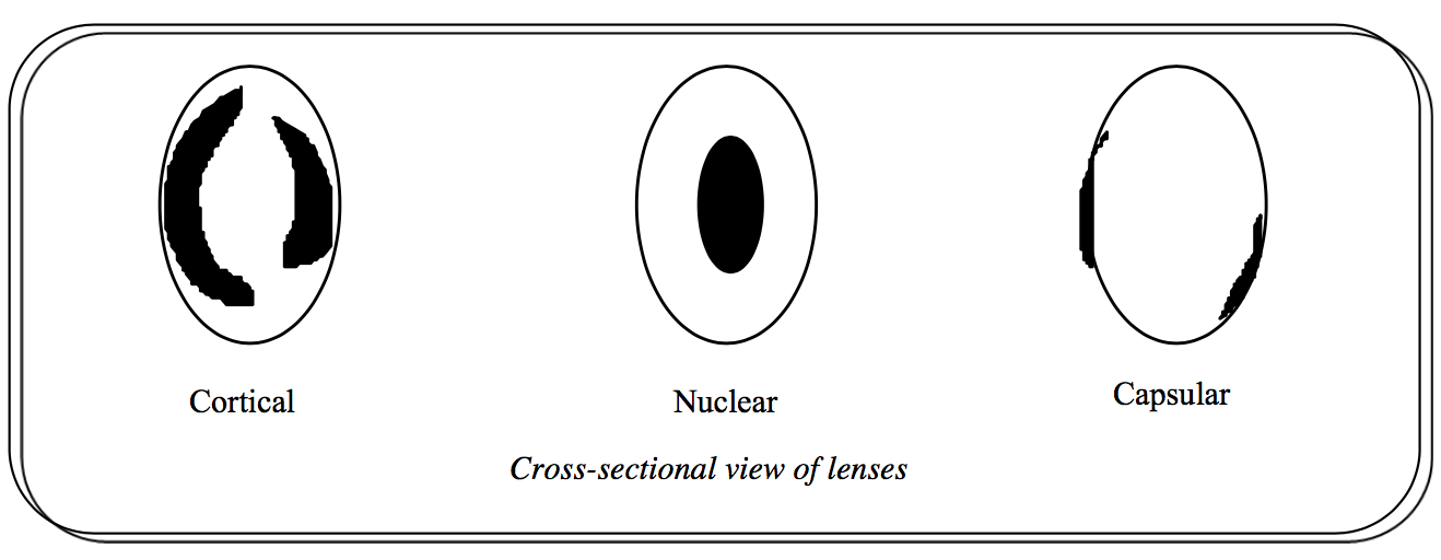
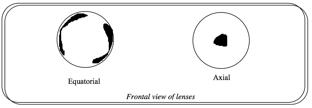
Description based on what part of the lens is involved.
Cortical, nuclear, capsular
Equatorial, axial
# Treatment
Surgery is the only effective means of treating cataracts.
Not all dogs with cataracts need to be treated. For example, an animal with an incipient equatorial opacity will have excellent vision, and as long as the opacity doesn’t progress its vision will remain excellent.
# Surgical techniques
Intracapsular cataract extraction (ICCE) - Through a large corneal incision and a pharmacologically dilated pupil, the ciliary zonules are cut and the entire lens is removed. Not recommended for routine cataract surgery; reserved primarily for removal of luxated lenses
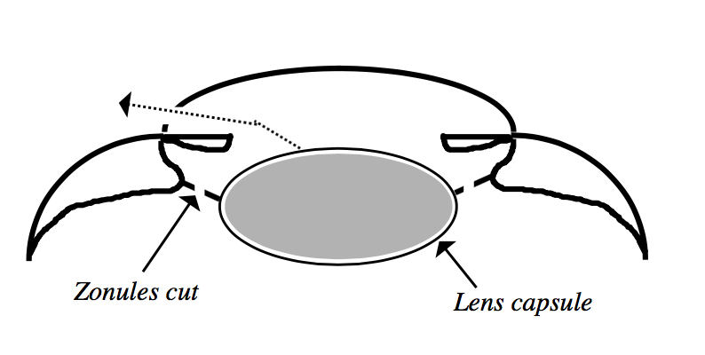
Extracapsular cataract extraction (ECCE) - Through a large corneal incision and pharmacologically dilated pupil a portion of the anterior lens capsule is removed and the lens nucleus and cortex are manually expressed through this “capsulotomy”. The equatorial and posterior portions of the lens capsule are left in place.
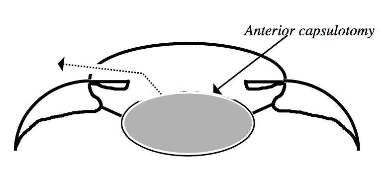
Phacoemulsification - A variation of ECCE. Through a small corneal incision, an anterior casulotomy is made and the nucleus is liquified using ultrasound (NOT LASER!!). The liquified nucleus and the cortex are then aspirated through the same small corneal incision (the cortex is soft enough that it can be aspirated without being ultrasounded). During the procedure the anterior chamber is irrigated so that the eye never collapses. A small handpiece simultaneously performs the ultrasound, irrigation, and aspiration.
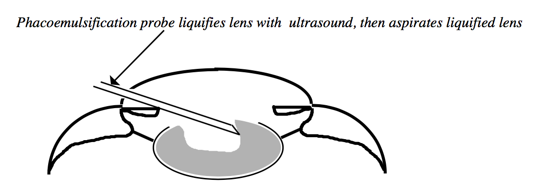
Your text has an excellent section on the details of phacoemulsification. While I encourage you to read this section, the detail provided is geared towards the ophthalmologist so don’t get bogged down in those details.
# Lens replacement
Prosthetic intraocular lenses (IOLS) are available for dogs. Without an IOL light rays will fall to focus behind the retina instead of on the retina — i.e., the dog will be hyperopic or “far-sighted”. The extent to which an IOL improves vision and quality of life in dogs is a hotly debated topic.

# When to refer for surgery
Phacoemulsification is currently the preferred method of cataract extraction. Surgical time is shorter, postoperative inflammation is reduced, and overall success rate is higher. However, surgery must be done fairly early because as the animal ages and the cataract becomes more mature, the lens becomes denser. If too dense, the ultrasound will not successfully dignify the nucleus.
It is advantageous to operate prior to the onset of hypermaturity because the uveitis that accompanies hypermaturity drastically reduces the success rate.
# Concurrent retinal disease
A fair percentage of dogs with cataracts also have a retinal disease called progressive retinal atrophy (PRA). PRA is an untreatable, blinding condition. Therefore doing cataract surgery in an animal with PRA will not improve vision. PRA can be diagnosed on retinal exam, but if a cataract is present it may be impossible to adequately visualize the retina. Fortunately we can assess retinal function even in the presence of a mature cataract with a test called an electroretinogram (ERG). An ERG is a standard part of all workups prior to cataract surgery.
# Nuclear Sclerosis
Definition: A senescent change in the lens that invariably occurs in dogs over 7 years of age. Occurs due to progressive lens fiber formation and internal compression of older fibers, especially in the lens nucleus. Sometimes called “lenticular sclerosis.”
Diagnosis: Seen as an overall haziness or grayness to the nucleus of the lens, but with sclerosis the lens is not opaque….i.e., light can still go through it. You can verify for yourself that light is going through the nucleus by either seeing a full fundic reflection or by being able to visualize the retina. The sclerosis (and therefore the haziness) becomes more pronounced with age.
Importance: Nuclear sclerosis itself does not interfere with vision, so the importance is in being able to differentiate it from nuclear cataract. In extremely old dogs (say, 16+ yrs) it can be very difficult to distinguish sclerosis from age-related nuclear cataract.
# Lens luxation
Definition: Dislocation of the lens from its normal position within the patellar fossa is referred to as subluxation (subtotal dislocation) or luxation (total dislocation, anterior and posterior), and is related to a pathologic alteration in the ciliary zonules from abnormal development, degeneration, rupture, tearing, or a combination of these factors.
Luxations are defined as “anterior” vs. “posterior” based on the anatomic relationship between the lens and the iris.
Posterior luxation: The lens remains behind the iris but is completely free of zonular attachments. An “aphakic crescent” (i.e., a crescent-shaped area within the pupil that lacks a lens) may be seen between the pupil and the equator of the lens. Sometimes the lens will fall all the way back in the vitreous and rest on the floor of the globe, and it will not be visible in the pupil.
Anterior luxation: The lens moves into the anterior chamber.
Subluxation: The lens remains behind the iris and is still partially held in place by zonules and only an aphakic crescent is seen.
# Diagnosis:
Observation of lens being out of position.
Tremors of the iris (iridodonesis) or of the lens (lenticulodenesis)
Focal axial corneal edema
Vitreous in the anterior chamber
Abnormally deep (if posteriorly luxated) or shallow (if anteriorly luxated) anterior chamber
# Etiologies
# Primary
Genetic
- Associated with a mutation in the ADAMTS17 gene
Occurs in terrier breeds (esp. the Jack Russell and the miniature bull terrier), Shar Pei and border collies, but can be seen in any breed
Always bilateral, although the two eyes almost never luxate at exactly the same time
Average age of onset is in the 3-6 yr old range.
Usually present as acutely painful, inflamed eyes.
# Secondary to:
Glaucoma
Will always be associated with buphthalmia
Therefore associated with chronic glaucoma
These are typically posterior subluxations
Hypermature cataract formation
Chronic uveitis
Aging
+/- Trauma
# Complications of lens luxation:
Glaucoma due to physical obstruction of the pupil or angle as well as other, less well defined alterations to the angle structure.
- Disturbingly, these animals may also have a concurrent POAG
Anterior uveitis
Corneal edema due to contact of the lens with the corneal endothelium
Retinal detachment
Posterior luxations / subluxations are often asymptomatic.
# Treatment for primary lens luxation:
SURGICAL EMERGENCY if acute!
Intracapsular cataract extraction (ICCE) - Through a large corneal incision, the entire lens is removed. Ideally, an artificial lens is sutured into the ciliary sulcus (i.e., area where iris meets ciliary body) after the luxated lens is removed.
Luxations secondary to chronic uveitis are not treated (surgically).
Posterior luxations are often treated conservatively, with pupillary constriction.
# Vitreous
Firm gel in the posterior segment filling the space between the lens and retina
# Congenital vascular anomalies of the vitreous
Persistent hyaloid artery – the hyaloid artery is a fetal structure that extends from the optic nerve head to the back of the lens. It should resorb late in gestation, but in some cases small pieces of it will persist into adulthood. In very rare circumstances such persistent fragments will be blood-filled. Generally considered a benign change.
Persistent tunica vasculosa lentis – The TVL is a spider-web like net around the embryonal lens that is fed by the HA. As with persistent HA, portions of it may persist into adulthood. Usually benign.
Persistent hyperplastic tunica vasculosa lentis / persistent hyperplastic primary vitreous. parts of the hyaloid system and primitive vitreous have become hyperplastic during early fetal development, combined with a subsequently incomplete regression. Seen in several breeds, most commonly the Dobie. Genetic. These dogs often also have developmental lens anomalies (microphakia, spherophakia, cataract, etc). Lens may actually have blood in it. Can cause visual disturbances, and surgical removal of the lens is usually necessitated but can be complicated by the potentially patent blood vessels.
# Acquired degenerative changes in the vitreous
Syneresis - Syneresis is a degenerative breakdown of the vitreous gel that separates its liquid from its solid components, resulting in liquefaction and development of fluid- filled cavities within the vitreous. There is some thought that this change may predispose to retinal detachment, especially in the Shih Tzu.
Asteroid hyalosis - Calcium soaps within the vitreous that appear as multifocal white suspended vitreal opacities (like glitter) that sway gently to and fro in the vitreal breeze as the dog moves the eyes back and forth. This is a fairly common aging change, and does not have an appreciable negative effect on the eye in most cases. I have seen cases in which asteroid is so dense that it appears to diminish vision. The “company line” on asteroid hyalosis is that it is a benign change that doesn’t indicate severe ocular pathology, but my personal opinion (also shared with your text) is that asteroid is seen more frequently in dogs with progressive retinal atrophy (which will study later in the term).
Synchysis scintillans - Particulate cholesterol crystals visible within a liquified vitreous; a degenerative change secondary to inflammation. If the vitreous is liquefied this has the appearance as a “snowstorm paperweight.”